2021 Best in Digital Drawing and Illustration – Amanda Cheung
Breakthrough
Medium: Digital drawing printed out onto paper flip book
The scientific concept behind my artwork features the mRNA COVID-19 vaccine (e.g. Pfizer and Moderna). Although this mRNA technique has been used primarily in research, the application of it to something as large-scale as a vaccine is what makes it a breakthrough.
I utilized a recreational mathematical construct called a tetraflexagon to illustrate how the steps of this mRNA technique works. A tetraflexagon is created from a strip of twelve connecting squares cut from a piece of square paper. The strip is then folded onto itself into a surface of four squares and can be manipulated to reveal a total of six different surfaces. For each face, I tried to come up with unique compositions and referred to biology textbooks to inspire my rendering. I made sure to colour code all the cell components, biological molecules, and backgrounds.
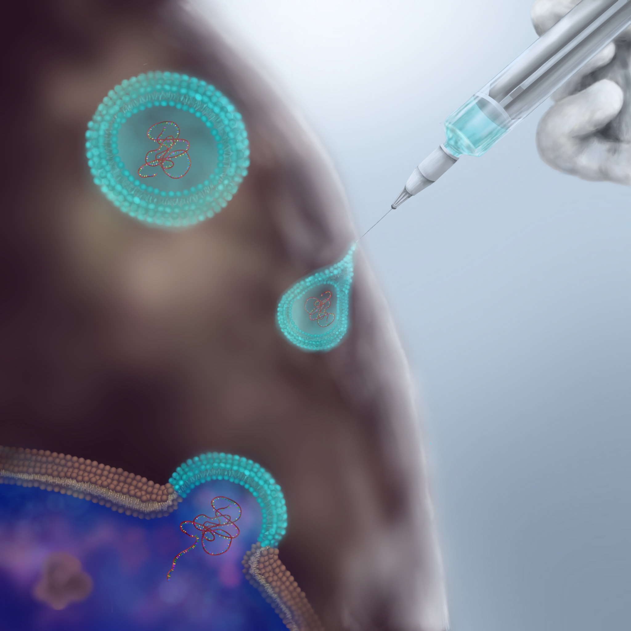
The first face shows the injection of the vaccine, introducing an mRNA sequence that contains instructions to create the “spike” proteins of the coronavirus. The mRNA is surrounded by a protective lipid nanoparticle layer, which has similar properties to the phospholipid bilayer in cell membranes. After injection, vaccine particles fuse to cells and release mRNA into the cytoplasm.
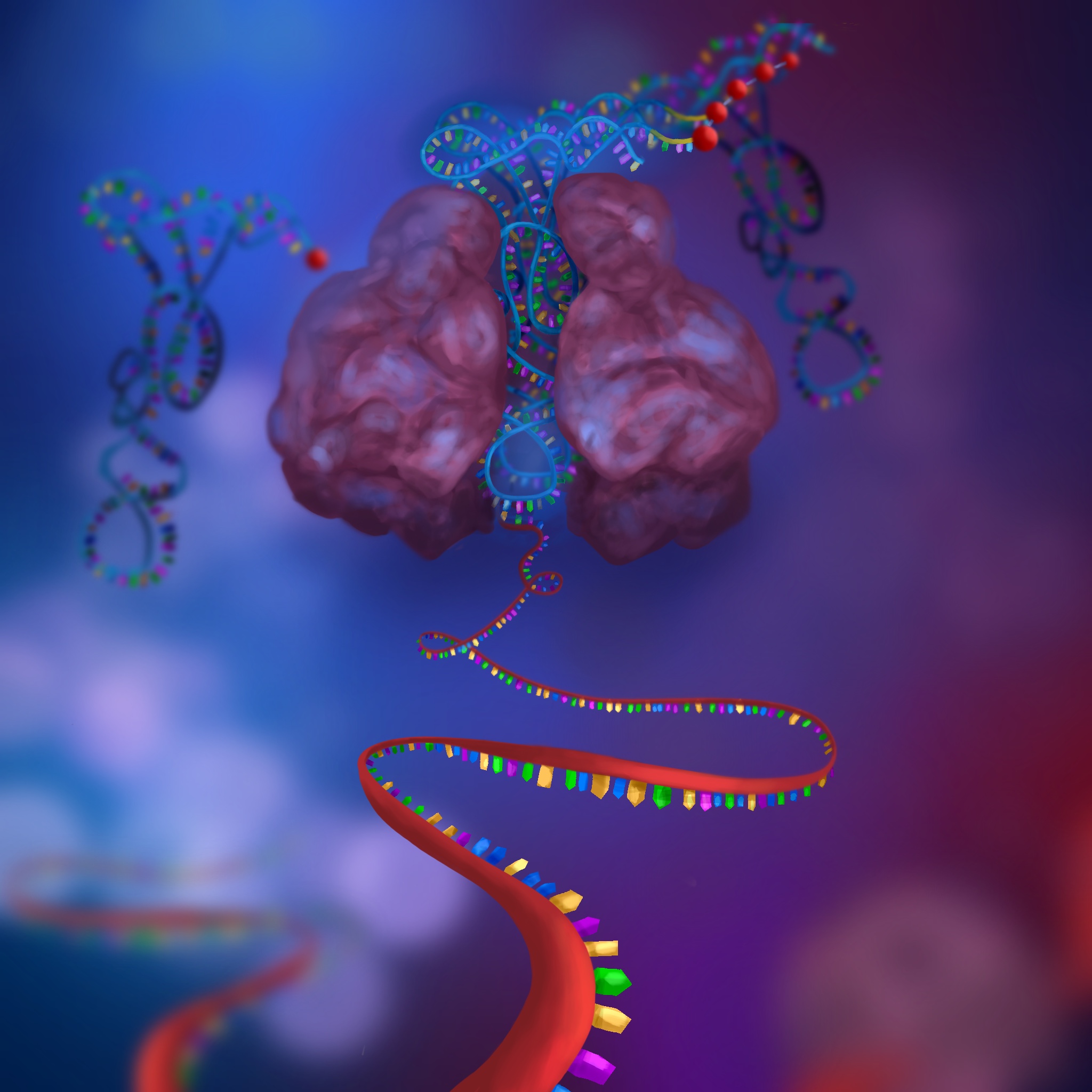
The second face shows the translation of mRNA into a spike protein. I showed how another molecule — tRNA — delivers anticodons that are complementary to the sequence on the mRNA and how it codes for a specific amino acid, which accumulates into a protein chain. The leftover mRNA is permanently degraded by enzymes.
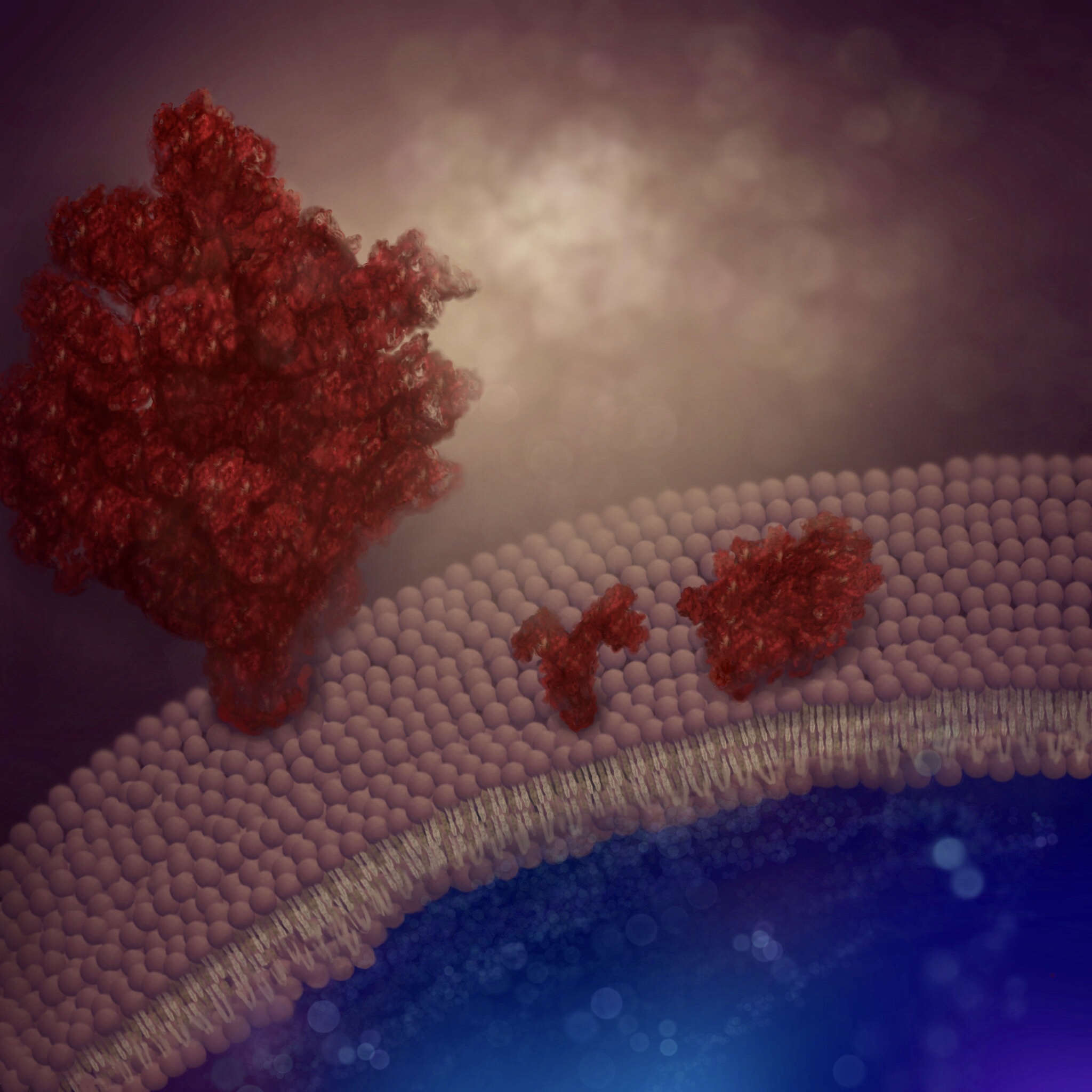
The third face shows the newly synthesised spike proteins and fragments on the surface of the cell. For simplicity, I left out the extraneous cell surface proteins that would usually be present.
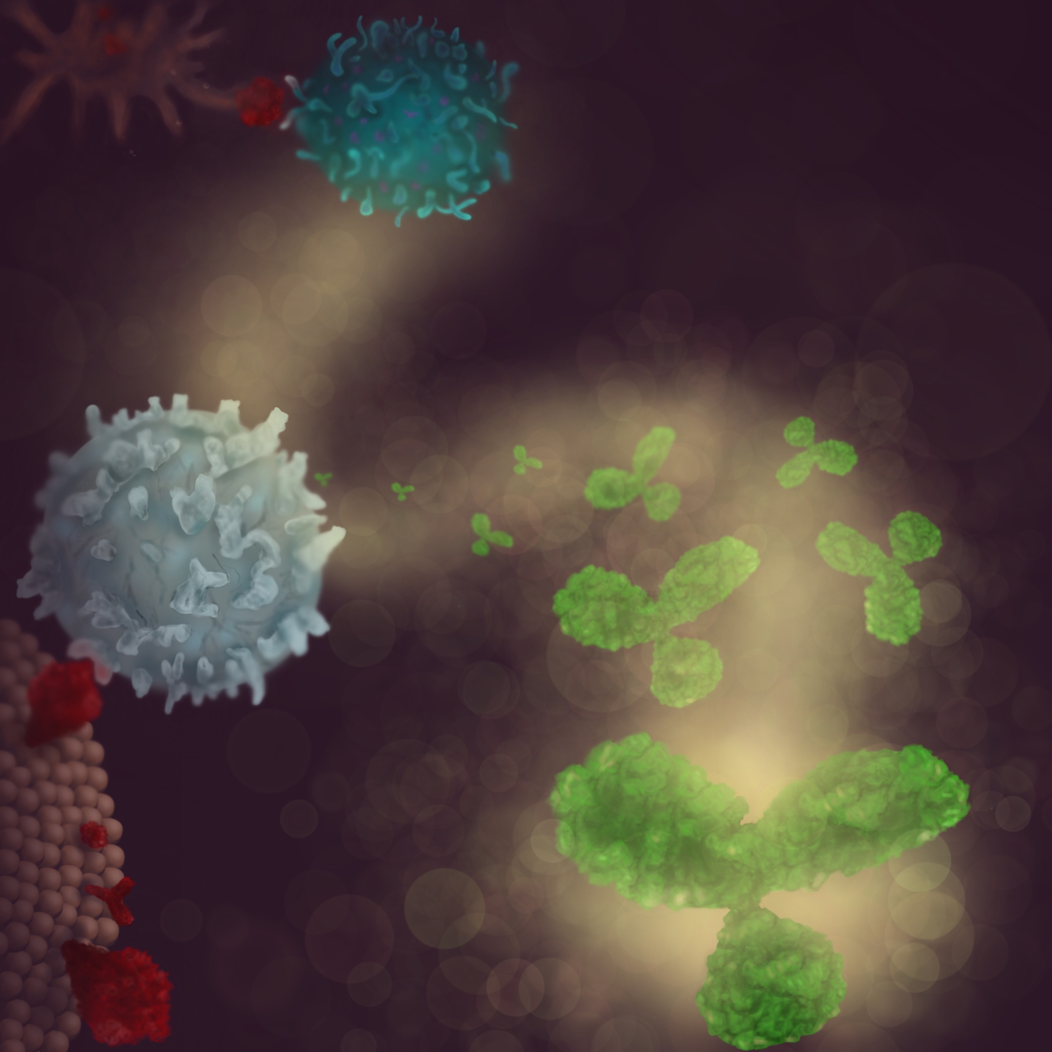
The fourth face shows an activation pathway. When a vaccinated cell dies, an antigen-presenting (dendritic) cell sweeps up protein fragments from the debris and presents them to our helper T cells. Other immune cells (B cells) lock onto spike proteins. This action causes helper T cells to activate B cells and secrete antibodies that are specialised in targeting the spike protein.
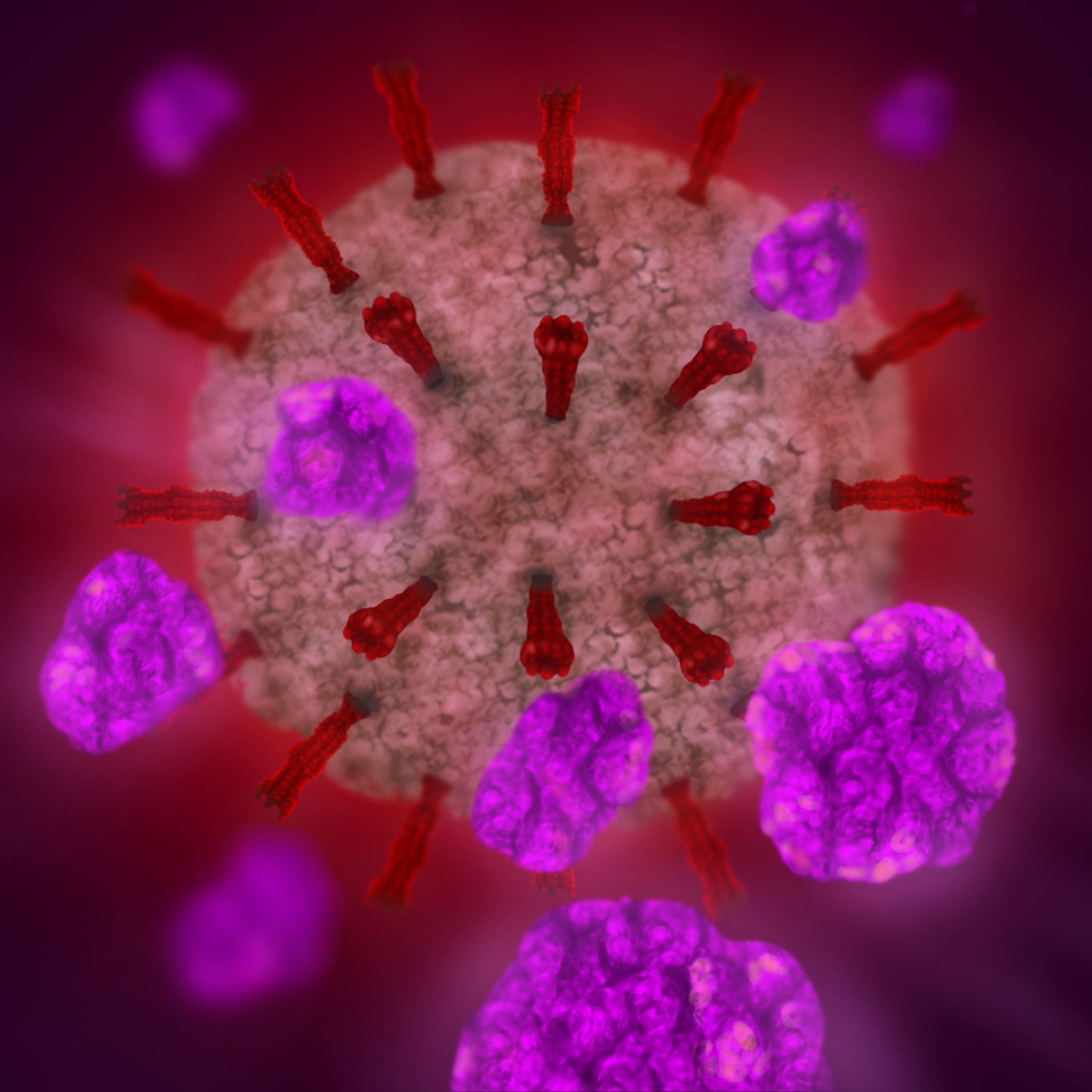
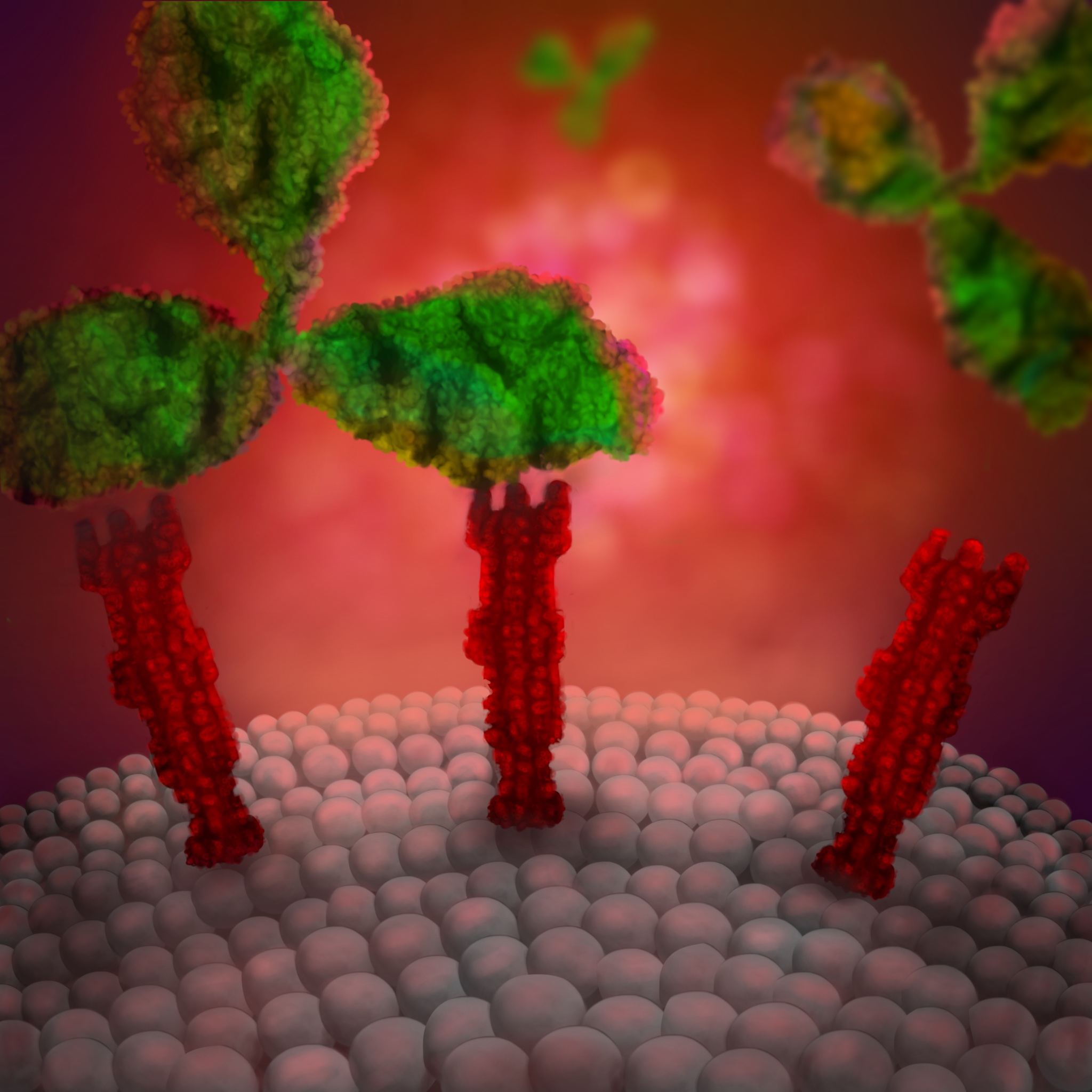
After going back from face 4 to 2, I demonstrated what occurs if a coronavirus enters the body. The fifth face shows the antibodies blocking the coronavirus proteins from attaching to other cells and marking the virus for destruction.
The last face shows activated killer T cells destroying an infected cell. A finding by research scientist Wang Nianshuang shows how coronavirus spikes are able to lock onto cell surface proteins in a process called membrane fusion. The coronavirus protein changes from a small mushroom shape (from when it first makes contact with a human cell) to a rod-like form that initiates entry and takes over the cell. The sequence of the stabilised, pre-fusion spike protein was incorporated into the vaccine.
As someone with a penchant for biology, I’m deeply interested in the inner workings regarding the coronavirus and the vaccine. It is breakthroughs like this which fuel my aspirations to go into medicine.
http://www.scmp.com/news/china/science/article/3116352/chinese-scientists-spike-protein-research-paves- way-covid-19
