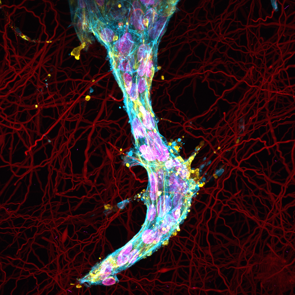
2021 People’s Choice Winner; Best in Photography – Alexander Zarouk

Metastasis
Medium: Digital Photography
Artistic Components
Immunostaining is the process of tagging specific proteins of a biological sample with a fluorescent antibody. By using different fluorescent tags, multiple proteins can be imaged at once and combined to form an image. This image was acquired with a Zeiss LSM 800 laser scanning confocal microscope that can image these fluorescent tags at subcellular resolution. Before the image is acquired, the microscope’s individual color channels must be tuned to reduce overexposure and the minimum and maximum focal distance are set. As the microscope acquires a stack of images taken within the range of focal lengths within a region of the hydrogel sample, the images are combined together to form a maximum projection that serves as a visual representation of the spheroid outgrowth. This is a direct parallel to a photographer’s use of low exposure shots to take captivating images of light trails at night. Through quantification of the intensity and spatial expression of these proteins, we can better understand cellular processes underlying metastasis.
Scientific Components
EMT (epithelial-mesenchymal transition) is the activation of cellular machinery that promote cancer cell invasion in the microenvironment of a primary solid tumor. EMT affects migratory behavior prior to and during metastasis through the loss of adhesion proteins on the cell surface. By breaking free from the primary tumor body, the cells now have metastatic potential to remodel the surrounding stroma. Due to the environmental stresses placed on a tumor, most cells of malignant potential will undergo EMT to migrate to a more suitable environment or confer resistance to cancer treatments.
Localized vimentin around an epithelial cell’s nuclei is indicative of the transition to a mesenchymal phenotype. It is the coexpression of both epithelial and mesenchymal markers that makes EMT a migratory driver of cell metastasis. As cells collectively move in an environment constituting collagen gel and its fiber matrix, the expression of EMT markers change. The fibers serve as contact-driven cues for cells to move away from their spheroid body, as occurs physiologically in breast stroma. Cells lose expression of their cell-cell contacts (e-cadherins) and serve to protect their nuclei through caging of vimentin as they navigate. Using a synthetic hydrogel system capable of replicating certain in-vivo aspects, the Baker Lab has been able to recapitulate EMT in a highly controllable manner through the tuning of fiber density, hydrogel stiffness, and other mechanical cues.
