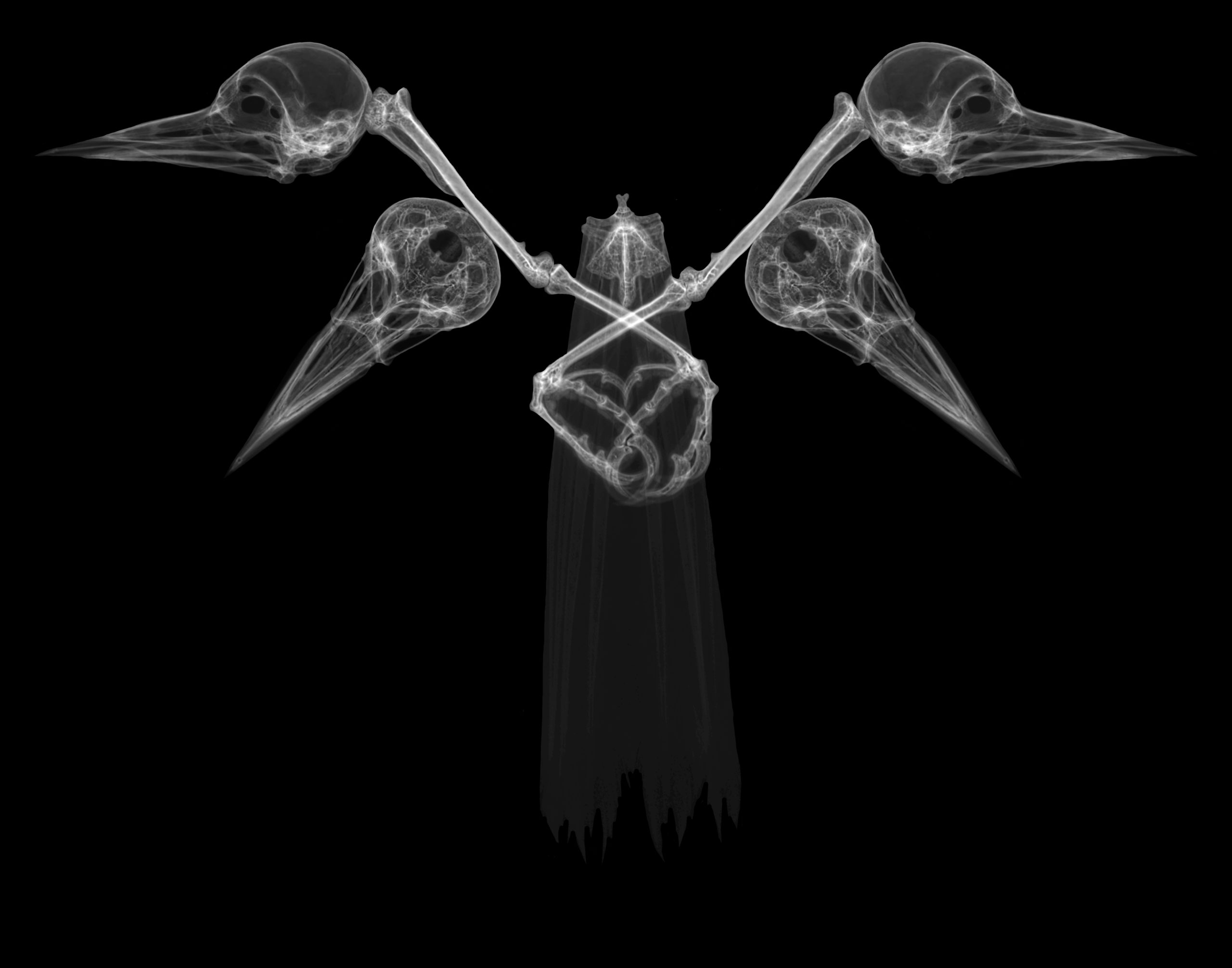
2020 Honorable Mention – Stephanie Francalancia

Protective Skulls
As humans, we must look to nature to understand the world around us. Scientists, even more interestingly, research nature and other organisms to find solutions to common problems humans encounter. Recently, I had the opportunity to work alongside researchers at the U of M Natural History Museum Research Center studying woodpeckers and the mechanisms by which the complex machinery of woodpeckers’ skulls prevent them from acquiring brain injuries as they peck wood. In my work, I decided to X-ray the woodpeckers’ skulls to create a piece that not only highlights these intricate brain structures but showcases them in an environment where viewers would not initially think of scientific research. Using the high-resolution images from the X-ray machine, I created a necklace-type structure with Photoshop to decontextualize the important studies of woodpecker physiology and make them feel familiar for audiences who are not usually concerned with woodpeckers, science, or research.
X-rays are typically used to diagnose brain injury and other abnormalities in humans. They function using the scattering of high energy photons toward the body. Since these photons have a shorter wavelength than visible light, they travel at a higher energy to be reflected or absorbed. Calcium atoms in bones and skeletal structures are large enough to absorb these photons, whereas the small size of atoms in tissue causes the photons to reflect. Thus, when photons are passed through the body onto a negative film, bones do not let the light photons shine through, and present as a lighter area. As depicted in this piece, X-rays are capable of demonstrating extreme detail. Whether for scientific research or artistic purposes, X-ray is an incredibly important and vital technology in modern medicine.
The clear resolution that the X-ray is able to obtain showcases all the minuscule yet integral bones that are a part of the woodpecker skull. For example, the hyoid bone in humans is a small and insignificant part of the neck. However, even this tiny bone in woodpeckers can be seen through X-rays to start in the beak and wrap all the way around the back of the skull. It does this in order to dissipate energy and force due to pecking laterally around the brain so as to not induce damage to the woodpecker’s cortex. The limbs depicted are also of importance to the bird as their sharp long claws allow them to hold onto the sides of trees and their strong tailbone helps with balance. Most importantly, these structures aid in redistributing the pecking force from the beak and skull back down into the bark of the tree.
Although this piece looks “intriguingly weird,” as described by my art professor, it also withholds invaluable information about how we can utilize nature, specifically woodpeckers, to potentially solve problems surrounding brain injury and impact.
