1st Year, LSA

Graphite, Ballpoint Pen, Watercolor
Abstract
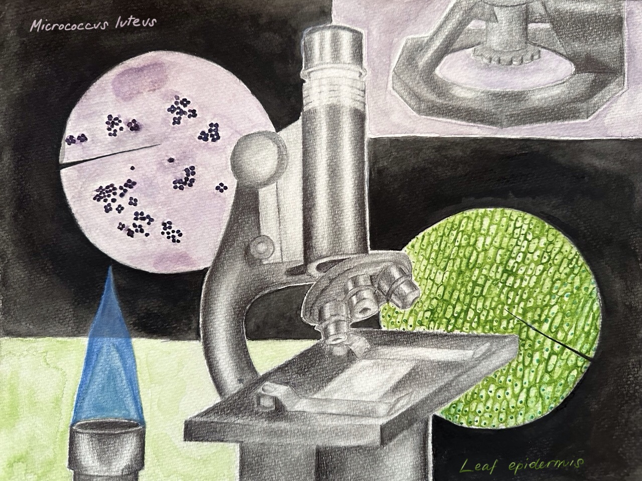
This mixed-media work visually represents the contrast between the abstract nature of living organisms and the industrial methods with which humans have used to explore and understand them. Using graphite, ballpoint pen, and simple, highlighter-like shades of watercolor, the work feels like the mind of a student studying this topic. That was my goal, to communicate how learning about biology felt like to me. As a consequence, this project allowed me to display the way that microbiology manifested in my mind.
Research for this piece preceded every other aspect of artistic planning, because it was the inspiration in the first place. I noticed the importance of attention to detail in order to identify specific structures under the microscope. Pathology and microbiology, in my opinion, are some of the most visually-oriented fields of science. I believe that I would not have the same level of passion for art had I not been involved in these subjects, and vice versa.
When choosing materials to use, I was interested in the ability to layer and build values gradually while maintaining vibrancy. I looked towards watercolor, as it seemed like an interesting challenge, especially since I hadn’t seriously practiced it before. This came with a lot of trial and error to find what techniques were best-suited for my skill set and purposes.
I worked with Micrococcus luteus and was able to see its growth under the microscope. I learned about its particular arrangement and coloration, which is determined by its absorption of gram stain. This bacteria is gram positive, meaning it has an outer peptidoglycan layer that allows the dye to permeate. This is in contrast to gram-negative bacteria, which has a fatty, glycolipid outer layer that acts as a wall. As for the other sample, my teacher provided me with a slide of pre-prepared leaf epidermis. Recreating its vibrant yet translucent earthy green required considerable practice with watercolor and shade matching. Nature is profoundly precise, and I found that demonstrating this precision was the most challenging aspect of creating this work.
The subject of this piece, the microscope, was symbolic of my experience in the lab. I kept it monochrome with graphite and ballpoint pen to separate it from the background and, in a way, create a comparison between the abstract art of nature and the meticulous construction of lab supplies. I used the same thinking when I kept the bunsen burner monochrome as well, apart from the organically-shaped flame. In order to see the true arrangement of the bacteria, the microscope was set to 40x, which is the second-highest magnification. This was not the case for the leaf epidermis, as I kept it on only 10x magnification to show the collection of plant cells. With this piece, I often elected to do what looked and felt best to me, as only so many creative liberties can be taken when attempting to demonstrate biology accurately. This is also why I chose to keep the composition geometric and rigid, similar to textbooks and lab manuscripts.
